


Characterization of the Curcuminoids Fingerprint Profile in Curcuma and Zingiber Genera by TLC – Digital Image Analysis
Abstract
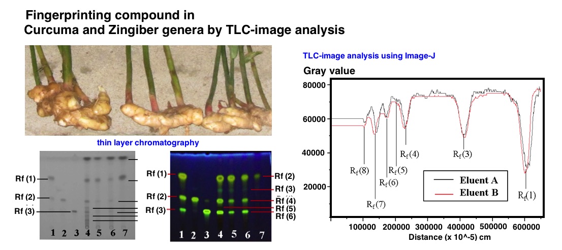
Curcuma longa, C. xanthorrhiza, C. heyneana, and Zingiber cassumunar contain high curcuminoid and have relatively the similar yellow color. Therefore, that they are potentially adulterated and difficult to differentiate in the form of powder. Hence, it is necessary to characterize the fingerprints compound profile by a simple and rapid method. This research aims to determine fingerprint compound profile of curcuminoid using Thin Layer Chromatography (TLC) and digital image analysis. The result of the research identified that the fingerprint compound profile of curcuminoid on the four rhizomes was obtained by TLC method using silica gel 60 GF254 as the stationary phase, chloroform:dichloromethane:methanol (13:6:1) as the mobile phase, and observation under UV 254 nm light and citroborate reagent. Thereafter, the digital image analysis was carried out using Image J software according to the gray value and % of RGB (red-green-blue) value. Based on gray value and % of RGB, both Curcuma and Zingiber genera were differentiated through curcumin compound (Rf 0.63), demethoxycurcumin (Rf 0.34), bisdemethoxycurcumin (Rf 0.21). The profile of fingerprint compound on Curcuma longa, C. xanthorrhiza, C. heyneana, and Zingiber cassumunar was differentiated through Rf 0.26; Rf 0.17; and Rf 0.10.
References
[1] Rafi, M., Rohaeti, E., Miftahudin, A., Darusman, L.K., Indones. J. Chem., 2011, 11 (1), 71–74
[2] Departemen Kesehatan Republik Indonesia, Farmakope Herbal Indonesia Edisi I, 2008, Departemen Kesehatan Republik Indonesia, Jakarta
[3] Nihayati, Ellis, Tatik Wardiyati, Rurini Retnowati, and Soemarno, Agrivita J. Agri. Sci., 2013, 35 (3), 218-226.
[4] Wonorahardjo, Surjani, Metode-Metode Pemisahan Kimia, 2016, Indeks Akademia, Jakarta
[5] Paramasivam, M., Poi, R., Banerjee, H., Bandyopadhyay, A., Food Chem., 2009, 113 (2), 640–644.
[6] Revathy, S., S. Elumalai, Merina Benny, and Benny Antony, J. Exp. Sci., 2011, 2(7), 21-25.
[7] Sirait, Midian, Penuntun Fitokimia dalam Farmasi, 2007, Penerbit ITB, Bandung.
[8] Tie-xin, Tang and Wu Hong, An Image Analysis System for Thin-Layer Chromatography Quantification and Its Validation, J. Chromatogr. Sci., 2008, 46 (6), 560-564.
[9] Firdaus, M. Lutfi, Wiwit Alwi, Ferli Trinoveldi, Iman Rahayu, Lena Rahmidar, Kancono Warsito, Procedia Environ. Sci., 2014, 20, 298-304.
[10] Rueden, C. T., Schindelin, J., Hiner, M.C., Dezonia, B.E., Walter, A.E., Arena, E.T. and Eliceiri, K.W., BMC Bioinformatics, 18(1), 529
[11] Ferreira, Tiago and Wayne Rasband, The Image J User Guide, http:// imagej.nih.gov/ij/docs/user-guide.pdf, date of access at 1 February 2019
[12] Priyadarsini, Kavirayani Indira. Moleculs, 2014, 19 (12), 20091-20112.
Refbacks
- There are currently no refbacks.

This work is licensed under a Creative Commons Attribution-NonCommercial 4.0 International License.