


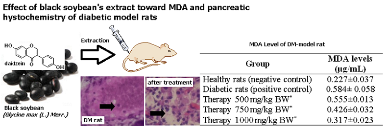
MDA and Histologic Profile of Pancreatic Diabetic-Rats Model Administered With Extract of Glycine max (L.) Merr.
Abstract
References
[1] American Diabetes Association, Diabetes Care, 2010, 33 (1), S62–S69.
[2] K. G. M. M. Alberti and P. ft Zimmet, Diabet. Med., 1998, 15 (7), 539–553.
[3] B. Lukiati, A. Aulanni’am, and W. Darmanto, Int. J. Basic Appl. Sci., 2012, 12 (2), 22–30.
[4] D. Del Rio, A. J. Stewart, and N. Pellegrini, Nutr. Metab. Cardiovasc. Dis., 2005, 15 (4), 316–328.
[5] R. Mateos, L. Goya, and L. Bravo, J. Chromatogr. B, 2004, 805 (1), 33–39.
[6] V. Peddireddy, B. Siva Prasad, S. D. Gundimeda, P. R. Penagaluru, and H. P. Mundluru, Biomarkers, 2012, 17 (3), 261–268.
[7] T. Widyawati, W. W. Purnawan, I. J. Atangwho, N. A. Yusoff, M. Ahmad, and M. Z. Asmawi, Int. J. Pharm. Sci. Res., 2015, 6 (4), 1698.
[8] A. Saifudin, T. Usia, S. AbLallo, H. Morita, K. Tanaka, and Y. Tezuka, Asian Pac. J. Trop. Biomed., 2016, 6 (1), 38–43.
[9] A. Saifudin, K. Tanaka, S. Kadota, and Y. Tezuka, J. Nat. Prod., 2013, 76 (2), 223–229.
[10] H. Y. Kil, E. S. Seong, B. K. Ghimire, I.-M. Chung, S. S. Kwon, E. J. Goh, K. Heo, M. J. Kim, J. D. Lim, and D. Lee, Food Chem., 2009, 115 (4), 1234–1239.
[11] V. K. Thirumalairaj, P. A. Anitha, G. Durairaj, S. G. Menon, and L. Shanmugaasokan, J. Appl. Pharm. Sci. Vol, 2014, 4 (12), 026–029.
[12] Y. Sireesha, R. B. Kasetti, S. A. Nabi, S. Swapna, and C. Apparao, Pathophysiology, 2011, 18 (2), 159–164.
[13] J. Zhang, E. Tatsumi, J. Fan, and L. Li, Int. J. Food Sci. Technol., 2007, 42 (3), 263–268.
[14] F. Fetriyuna, Int. J. Adv. Sci. Eng. Inf. Technol., 2015, 5 (1), 44–46.
[15] C. U. Zanetta, B. Waluyo, M. Rachmadi, and A. Karuniawan, Energy Procedia, 2015, 65, 29–35.
[16] L. P. Gina, C. Mahdi, and A. Aulanni’am, J. Pure Appl. Chem. Res., 2014, 3 (3), 131–137.
[17] B. Xu and S. K. Chang, J. Agric. Food Chem., 2008, 56 (16), 7165–7175.
[18] M.-G. Choung, I.-Y. Baek, S.-T. Kang, W.-Y. Han, D.-C. Shin, H.-P. Moon, and K.-H. Kang, J. Agric. Food Chem., 2001, 49 (12), 5848–5851.
[19] J. H. Lee, N. S. Kang, S.-O. Shin, S.-H. Shin, S.-G. Lim, D.-Y. Suh, I.-Y. Baek, K.-Y. Park, and T. J. Ha, Food Chem., 2009, 112 (1), 226–231.
[20] T. Tsuda, Y. Kato, and T. Osawa, FEBS Lett., 2000, 484 (3), 207–210.
[21] J.-M. Kong, L.-S. Chia, N.-K. Goh, T.-F. Chia, and R. Brouillard, Phytochemistry, 2003, 64 (5), 923–933.
[22] M. Masruri, M. Lutfillah, A. Sumaryanto, R. Retnowati, and A. ’am Aulanni’am, J. Trop. Life Sci., 2014, 4 (3), 161–165.
[23] J. Lee, R. W. Durst, and R. E. Wrolstad, J. AOAC Int., 2005, 88 (5), 1269–1278.
Refbacks
- There are currently no refbacks.